Herniated Thoracic Disc
A herniated disc occurs when the intervertebral disc's annulus (the outer fibers) is damaged and the soft inner material of the nucleus pulposus ruptures out of its normal space. If the annulus tears near the spinal canal, the nucleus pulposus material can push into the spinal canal. There is very little extra space around the spinal cord in the thoracic area. So when a herniated disc occurs in the mid back it can be extremely serious. In severe cases, the pressure on the spinal cord can lead to paralysis below the waist. Fortunately, herniated discs are not nearly as common in the thoracic spine as in the lumbar spine.
Learn about herniated thoracic disc including
- what causes a herniated thoracic disc
- how a diagnosis is made
- what treatment options are available
- what complications can arise
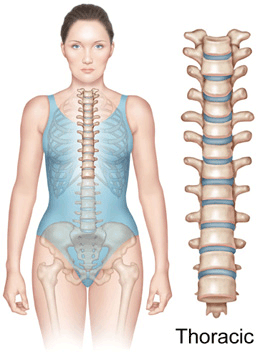
Anatomy
In order to understand your symptoms and treatment choices, it is helpful to start with a basic understanding of the anatomy of the mid back. Become familiar with the various parts that make up the thoracic spine and how they work together.
Learn more about the anatomy of the thoracic spine.
The intervertebral discs are the shock-absorbing cushions between each vertebra of your spine. There is one disc between each vertebra. Each disc has a strong outer ring of fibers, called the annulus, and a soft, jelly-like center, called the nucleus pulposus.
The annulus is the disc's outer layer and the strongest area of the disc. The annulus is actually a strong ligament that helps connect each vertebra together. The nucleus in the center of the disc serves as the main shock absorber.
A herniated disc occurs when the intervertebral disc's outer fibers (the annulus) are damaged and the soft inner material of the nucleus pulposus ruptures out of its normal space. If the annulus tears near the spinal canal, the nucleus pulposus material can push into the spinal canal.
Causes

Herniated discs can occur in children, although it is rare. A true herniated nucleus pulposus is most common in young and middle-aged adults and generally occurs in the low back. Disc herniations in the thoracic spine mostly affect people between age 40 and 60. In older folks, the degenerative changes that occur in the spine with aging make it less likely for them to suffer a true herniated disc.
Discs can rupture suddenly because of too much pressure all at once. For example, falling from a ladder and landing in a sitting position can cause a great amount of force through the spine. If the force is strong enough, either a vertebra can break or a disc can rupture. Bending places high forces on the discs between each vertebra. If you bend and try to lift something that is too heavy, the force can cause a disc to rupture.
Discs can also rupture from a small amount of force, usually due to weakening of the annulus from repeated injuries that add up over time. As the annulus becomes weaker, at some point lifting or bending causes too much pressure across the disc. The weakened disc ruptures while doing something that five years earlier would not have caused a problem. This is due to the effects of aging on the spine-the most common reason for a disc herniation in the thoracic spine.
The material that has ruptured into the spinal canal from the nucleus pulposus can cause pressure on the nerves in the spinal canal. There is also some evidence that the nucleus pulposus material causes a chemical irritation of the nerve roots. Both the pressure on the nerve root and the chemical irritation can lead to problems with how the nerve root functions. The combination of the two can cause pain, weakness, and numbness in the area of the body to which the nerve supplies sensation.
In the thoracic spine, the pressure can also affect the spinal cord. This is due to the fact that there is little extra space within the spinal canal of the thoracic spine. Too much pressure on the spinal cord can lead to paralysis from the waist down.
Symptoms
The first symptom of a thoracic disc herniation is usually pain. The pain is most often felt in the back, directly over the sore disc. Pain may also radiate around to the front of the chest. Pressure or irritation on the nerves in the thoracic area can also cause symptoms. Depending on which nerves are affected, a thoracic disc herniation can include pain that feels like it is coming from the heart, abdomen, or kidneys.
Herniated thoracic discs sometimes press against the spinal cord. When this happens, symptoms may include
- muscle weakness, numbness, or tingling in one or both legs
- increased reflexes in one or both legs that can cause spasticity
- changes in bladder or bowel function
- paralysis from the waist down
Diagnosis
History and Physical Exam
Diagnosing a herniated nucleus pulposus begins with a complete history of the problem and a physical exam. Your doctor will want to make sure that you are aware when you have to urinate or have a bowel movement. If there is a problem, it could indicate that a herniated disc in the thoracic spine is pushing against the spinal cord.
Diagnostic Tests
X-rays
The doctor may suggest taking X-rays of your mid back. Regular X-rays can't show a herniated disc, but they can give your doctor an idea of how much wear and tear is present in the spine. X-rays can also show a disc that has become calcified, as often happens to a herniated thoracic disc. If part of the calcified disc appears to be pointing into the spinal cord, it's a good indication the thoracic disc is herniated.
MRI
The most common test to diagnose a thoracic herniated disc is the MRI scan. This test is painless and very accurate. It is usually the preferred test to do (after X-rays) if a herniated thoracic disc is suspected.
CT Scan
Sometimes, the X-ray and MRI do not tell the whole story. Other tests may be suggested. A myelogram, usually combined with a CT scan, may be necessary to give as much information as possible.
Treatment Options
A herniated disc does not necessarily mean that you will need to undergo surgery. The treatment of a herniated disc depends on the symptoms. If the symptoms are getting better, your doctor may suggest watching and waiting to see if they go away. If they are getting steadily worse, your doctor may be more likely to suggest surgery. Many people, who initially have problems due to a herniated disc, find their symptoms completely resolve over several weeks or months.
Conservative Treatment
Observation
You may not need any treatment other than watching to make sure that the problem does not progress. If the pain is bearable and symptoms from nerve or spinal cord pressure are not getting worse, your doctor may just want to watch and wait.
Rest
If the pain is more severe, it may be necessary to take a few days off from work and decrease your activities. Your doctor may also prescribe a back brace to help limit movement around the injured disc. After several days, you should begin to get moving. Start with a gentle walking program and increase the distance you walk each day.
Pain medications
Depending on the severity of your pain, medications can be used to help control it. Over-the-counter pain relievers, such as ibuprofen, Tylenol(tm), and some of the newer anti-inflammatory medications, may be helpful.
If these types of medications do not control the pain, your doctor may prescribe stronger pain pills-narcotic or non-narcotic pain medications. Narcotic pain medications are very strong but also very addictive. Non-narcotic pain medications are less addictive, but are somewhat less effective than narcotics. Most physicians do not like to prescribe narcotics for more than a few days or weeks. Learn more about medications used to treat back pain.
Physical Therapy
If your condition is causing only mild symptoms and does not appear to be getting worse, your doctor may have you work with a physical therapist. A well-rounded rehabilitation program assists in calming pain and inflammation, improving your mobility and strength, and helping you do your daily activities with greater ease and ability.
In very mild cases, physical therapy offers ways to control symptoms and enable you to improve without surgery. Treatments focus on improving mobility and posture. Therapy sessions may be scheduled two to three times each week for up to six weeks.
The goals of physical therapy are to help you
- learn ways to manage your condition and control the symptoms
- learn correct posture and body movements to reduce back strain
- identify symptoms of thoracic herniation that require medical attention
- learn ways to manage your condition
Learn more about spinal rehabilitation.
Surgical Treatment
Laminotomy and Discectomy
The traditional way of surgically treating a herniated thoracic disc used to be to perform laminotomy and discectomy. The term laminotomy means "make an opening in the lamina", and the term discectomy means "remove the disc." The purpose of taking out a herniated thoracic disc was to decompress the spinal cord or spinal nerves. But nerve problems that occurred with this traditional method of decompression have led many doctors to discontinue this form of surgery for disc herniations in the thoracic spine.
Transthoracic Decompression
A new way to decompress the spinal cord or spinal nerves is a technique called transthoracic decompression. Operating from the patient's side, the doctor makes a small opening through the ribs and works on the spine through the chest cavity. A minimal amount of the vertebral body and problem disc are removed, taking pressure off the spinal cord. Fusion surgery is sometimes needed right afterward if a larger section of the vertebra has to be taken out.
Costotransversectomy
Pressure on the spinal cord from a herniated thoracic disc can also be effectively treated using a surgical procedure called costotransversectomy. The surgeon makes an incision through the back of the spine. The ends of one or more nearby ribs are removed where they join the spine. A section of the transverse process (the small bone on the side of the vertebra) is taken off. This forms an opening for the doctor to see the injured disc. The surgeon can then decompress the spinal cord by locating and removing the disc material that has ruptured into the spinal canal.
Video Assisted Thoracoscopy Surgery (VATS)
VATS is a new way to perform thoracic surgery. Only small incisions are required. The thoracoscope is a small T V camera that is passed through the chest cavity. Watching on a TV screen, the surgeon can see and treat the herniated disc. Because the incisions are small, most patients have an easier time recovering from the procedure.
Fusion
Fusion surgery joins two or more bones into one solid bone. The medical term for this procedure is arthrodesis. If surgery on the herniated thoracic disc requires removal of a large section of bone and disc material, the section of spine may become loose or unstable. When this happens it may be necessary to fuse the bones right above and below the unstable section.
Bone graft material is used to get the unstable bones to grow together. Rods, plates, and screws are commonly used to hold the bones in place so the bone graft heals. Learn more about spinal fusion.
Complications
Like all surgical procedures, operations on the back may have complications. Because the surgeon is operating around the spinal cord, back operations are always considered extremely delicate and potentially dangerous. Take time to review the risks associated with thoracic spine surgery with your doctor. Make sure you are comfortable with both the risks and the benefits of the procedure planned for your treatment. Learn more about possible complications of spine surgery.