Anatomy of the Spine
Introduction
Your spine is one of the most important parts of your body. It gives your body structure and support. Without it you could not stand up or keep yourself upright. It allows you to move about freely and to bend with flexibility. The spine is also designed to protect your spinal cord. The spinal cord is a column of nerves that connects your brain to the rest of your body, allowing you to control your movements. Without a spinal cord you could not move any part of your body, and your organs could not function. Keeping your spine healthy is vital if you want to live an active life. Learn about the spine including:
- how the spine functions
- what differentiates the cervical, thoracic and lumbar spinal segments
- what important structures make up the spine
Anatomy
What exactly is the spine?
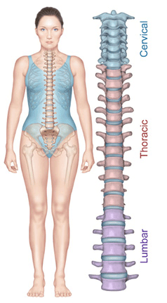
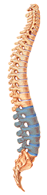
The spine is made up of 24 bones, called vertebrae. Ligaments and muscles connect these bones together to form the spinal column. The spinal column gives the body form and function. The spinal column holds and protects the spinal cord, which is a bundle of nerves that sends signals to other parts of the body. The many muscles that connect to the spine help support the upright posture of the spine and move the spine.
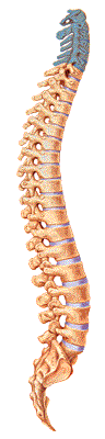
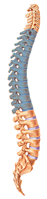
The spinal column has three main sections-the cervical spine, the thoracic spine, and the lumbar spine. The first seven vertebrae form the cervical spine. The mid back, called the thoracic spine, consists of 12 vertebrae. The lower portion of the spine, called the lumbar spine, is usually made up of five vertebrae. However some people have a sixth lumbar vertebra.
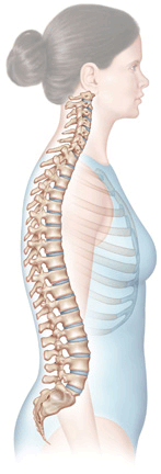
The normal spine has an "S"-like curve when looking at it from the side. This allows for an even distribution of weight. The "S" curve helps a healthy spine withstand all kinds of stress. The cervical spine curves slightly inward, the thoracic slightly outward, and the lumbar slightly inward. Even though the lower portion of your spine holds most of the body's weight, each segment relies upon the strength of the others to function properly.
Cervical Spine (Neck)
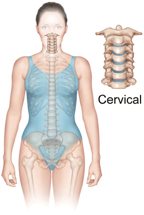
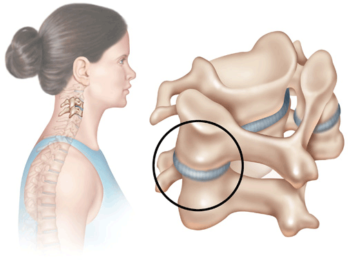
The cervical spine is made up of the first seven vertebrae in the spine. It starts just below the skull and ends just above the thoracic spine. The cervical spine has a lordotic curve, a backward "C"-shape-just like the lumbar spine. The cervical spine is much more mobile than both of the other spinal regions. Think about all the directions and angles you can turn your neck.
Unlike the rest of the spine, there are special openings in each vertebra in the cervical spine for arteries (blood vessels that carry blood away from the heart). The arteries that run through these openings bring blood to the brain.
Two vertebrae in the cervical spine, the atlas and the axis, differ from the other vertebrae because they are designed specifically for rotation. These two vertebrae are the reason your neck can move in so many directions.
The atlas is the first cervical vertebra-the one that sits between the skull and the rest of the spine. The atlas does not have a vertebral body, but it does have a thick forward (anterior) arch and a thin back (posterior) arch with two prominent sideways masses.
The atlas sits on top of the second cervical vertebra, the axis. The axis has a bony knob called the odontoid process, which sticks up through the hole in the atlas. Special ligaments between the atlas and the axis allow for a great deal of rotation. It is this special arrangement that allows the head to turn from side to side as far as it can.
The cervical spine is very flexible, but it is also very much at risk for injury from strong, sudden movements, such as whiplash-type injuries. This high risk of harm is due to the limited muscle support that exists in the cervical area, and the fact that this part of the spine has to support the weight of the head-an average of 15 pounds. This is a lot of weight for a small, thin set of bones and soft tissues to bear. Sudden, strong head movements can cause damage.
Thoracic Spine (Mid Back)
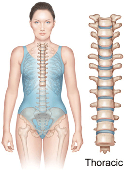
The thoracic spine is made up of the middle 12 vertebrae. These vertebrae connect to your ribs and form part of the back wall of the thorax (the ribcage area between the neck and the diaphragm). The thoracic spine's curve is kyphotic, a "C"-shaped curve with the opening of the "C" in the front. This part of the spine has very narrow, thin intervertebral discs. Rib connections and smaller discs in the thoracic spine limit the amount of spinal movement in the mid back compared to the lumbar or cervical parts of the spine. There is also less space inside the spinal canal.
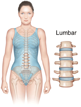
Lumbar Spine (Low Back)
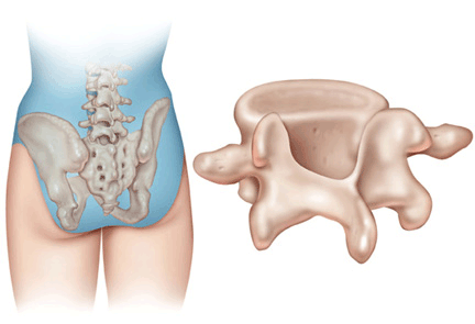
The lowest part of the spine is called the lumbar spine. This area usually has five vertebrae. However, sometimes people are born with a sixth vertebra in the lumbar region. The base of your spine (called the sacrum) is a group of specialized vertebrae that connects the spine to the pelvis. When one of the bones forms as a vertebra rather than part of the sacrum, it is called a transitional (or sixth) vertebra. This occurrence is not dangerous and does not appear to have any serious side effects.
The lumbar spine's shape has a lordotic curve-shaped like a backward "C". If you think of the spine as having an "S"-like shape, the lumbar region would be the bottom of the "S". The vertebrae in the lumbar spine area are the largest of the entire spine. The lumbar spinal canal is also larger than in the cervical or thoracic parts of the spine. The size of the lumbar spine allows for more space for nerves to move about.
Low back pain is a very common complaint for a simple reason. Since the lumbar spine is connected to your pelvis, this is where most of your weight bearing and body movement takes place. Typically this is where people tend to place too much pressure, such as when lifting up a heavy box, twisting to move a heavy load, or carrying a heavy object. These activities can cause repetitive injuries that can lead to damage to the parts of the lumbar spine.
Important Structures of the Spine
Your spine is made up of 24 small bones, called vertebrae. The vertebrae protect and support the spinal cord. They also bear the majority of the weight put upon your spine. Vertebrae, like all bones, have an outer shell, called cortical bone, which is hard and strong. The inside is made of a soft, spongy type of bone, called cancellous bone.
The vertebral body is the large, round portion of bone. Each vertebra is attached to a bony ring. When the vertebrae are stacked one on top of the other, the rings create a hollow tube for the spinal cord to pass through. Each vertebra is held to the others by groups of ligaments. Ligaments connect bones to bones; tendons connect muscles to bones. There are also tendons that fasten muscles to the vertebrae.
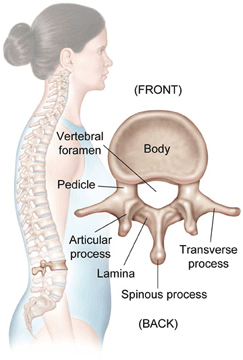
The bony ring attached to the vertebral body consists of several parts. The laminae extend from the body to cover the spinal canal, which is the hole in the center of the vertebra. The spinous process is the bony portion opposite the body of the vertebra. You feel this part if you run your hand down a person's back. There are two transverse processes (little bony bumps), where the back muscles attach to the vertebrae. The pedicle is a bony projection that connects the laminae to the vertebral body.

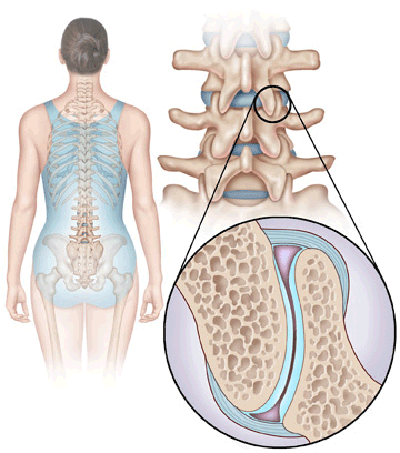
Between each vertebra is a soft, gel-like cushion, called an intervertebral disc. These flat, round "cushions" act like shock absorbers by helping absorb pressure. The discs prevent the bones from rubbing against each other.
Each disc has a strong outer ring of fibers called the annulus, and a soft, jelly-like center called the nucleus pulposus. The annulus is the strongest area of the disc. It helps keep the disc's center intact. The annulus is actually a strong ligament that connects each vertebra together.
The mushy nucleus of the disc serves as the main shock absorber. The nucleus is made up of tissue that is very moist because it has high water content. The water content is what helps the disc act like a shock absorber-somewhat like a waterbed mattress.
The spinal column has real joints (just like the knee, elbow, etc.) called facet joints. The facet joints link the vertebrae together and give them the flexibility to move against each other. The facets are the "bony knobs" that meet between each vertebra. There are two facet joints between each pair of vertebrae, one on each side. They extend and overlap each other to form a joint between the neighboring vertebra facet joint. The facet joints give the spine its flexibility.
The facet joints are synovial joints, structures that allow movement between two bones. The ends of the bones that make up a synovial joint are covered with articular cartilage, a slick spongy material that allows the bones to glide against one another without much friction. Synovial fluid inside the joint keeps the joint surfaces lubricated, like oil lubricates the parts of a machine. This fluid is contained inside the joint by the joint capsule, a watertight sac of soft tissue and ligaments that fully surrounds and encloses the joint.
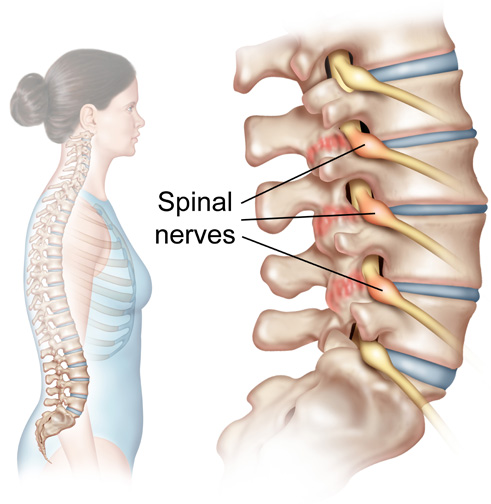
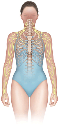
The spinal cord branches off into 31 pairs of nerve roots, which exit the spine through small openings on each side of the vertebra called neural foraminae. The two nerve roots in each pair go in opposite directions when traveling through the foraminae. One goes out the left foramina; the other goes out through the right foramina. The nerve root allows nerve signals to travel to and from your brain to the rest of your body
The spinal cord is a column of millions of nerve fibers that carries messages from your brain to the rest of your body. It extends from the brain to the area between the end of your first lumbar vertebra and top of your second lumbar vertebra. Each vertebra has a hole in the center, so when they stack on top of each other they form a hollow tube (spinal canal) that holds and protects the entire spinal cord and its nerve roots.
The spinal cord only goes down to the second lumbar vertebra. Below this level, the spinal canal contains a group of nerve fibers, called the caude equina. This group of nerves goes to the pelvis and lower limbs.
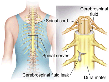
A protective membrane, called the dura mater covers the spinal cord. The dura mater forms a watertight sac around the spinal cord and the spinal nerves. Inside this sac, the spinal cord is surrounded by spinal fluid.
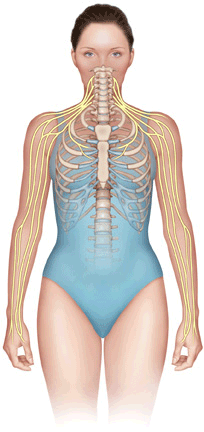
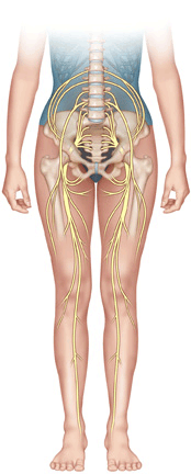
The nerve fibers in your spinal cord branch off to form pairs of nerve roots that travel through the small openings between your vertebrae. The nerves in each area of the spinal cord connect to specific parts of your body. This is why damage to the spinal cord can cause paralysis in certain areas and not others. It depends on which spinal nerves are affected. The nerves of the cervical spine go to the upper chest and arms. The nerves of the thoracic spine go to the chest and abdomen. The nerves of the lumbar spine reach to the legs, pelvis, bowel, and bladder. These nerves coordinate and control all the body's organs and parts, and allow you to control your muscles.
The nerves carry electrical signals back to the brain that allow you to feel sensations. If your body is being hurt in some way, your nerves signal the brain. Damage to the nerves themselves can cause pain, tingling, or numbness in the area where the nerve travels. Without nerve signals, your body would not be able to function.
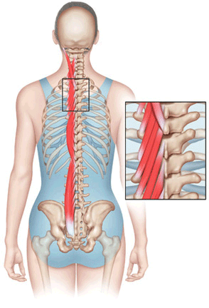
The muscles next to the spine are called the paraspinal muscles. They support the spine and provide the motor for movement of the spine. Joints allow flexibility, and muscles allow mobility. There are many small muscles in the back. Each controls some part of the total movement between the vertebrae and the rest of the skeleton. These muscles can be directly injured, such as when you have a pulled muscle or muscle strain. They can also cause problems indirectly, such as when they are in spasm after injury to other parts of the spine.
A muscle spasm is experienced when your muscle tightens up and will not relax. Spasms usually occur as a reflex (meaning that you cannot control the contraction). When any part of the spine is injured-including a disc, ligament, bone, or muscle-the muscles automatically go into spasm to reduce the motion around the area. This mechanism is designed to protect the injured area.
Muscles that are in spasm produce too much lactic acid, a waste product from the chemical reaction inside muscle cells. When muscles contract, the small blood vessels traveling through the muscles are pinched off (like a tube pinched between your thumb and finger), which causes a build up of lactic acid. If the muscle cells cannot relax and too much lactic acid builds up, it causes a painful burning sensation. The muscle relaxes as the blood vessels open up, and the lactic acid is eventually washed away by fresh blood flowing into the muscle.

Doctors sometimes look at a spinal segment to understand and explain how the whole spine works. A spinal segment is made up of two vertebrae attached together by ligaments, with a soft disc separating them. The facet joints fit between the two vertebrae, allowing for movement, and the neural foraminae between the vertebrae allow space for the nerve roots to travel freely from the spinal cord to the body. The spinal segment allows physicians to examine the repeating parts of the spinal column to understand what can go wrong with the various parts of the spine.