Cervical Laminectomy
Introduction

Learn about cervical laminectomy including
- how the cervical spine is affected
- why a laminectomy is performed
- what you can expect from this procedure including possible complications
- how rehabilitation can improve your results
Anatomy
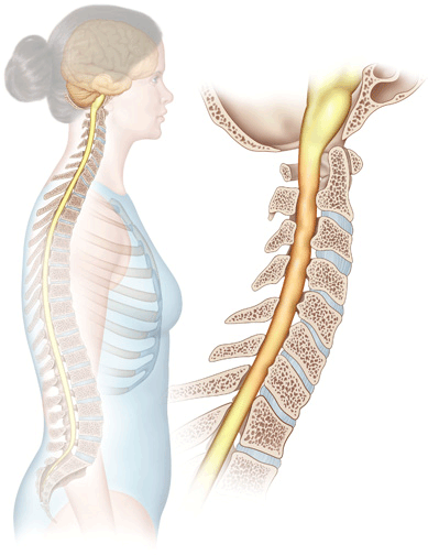
In order to understand your symptoms and treatment choices, it is helpful to start with a basic understanding of the anatomy of the neck. This includes becoming familiar with the various parts that make up the cervical spine and how they work together.
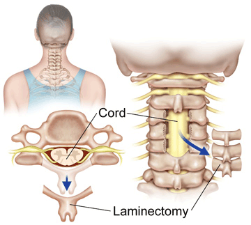
The bones of the spinal column protect the spinal cord. The vertebral body at each level of the spine protects the front of the spinal cord. The pedicle and lamina bones form a protective ring of bone that surrounds the sides and back of the spinal cord. The pedicles connect to the vertebral body. The lamina bones attach to the pedicles. The lamina bones cover the back surface of the spinal canal, forming a protective roof over the spinal cord.

Rationale
Bone spurs or a herniated disc can take up space inside the spinal canal and put pressure on the spinal cord. This condition is called spinal stenosis. If spinal stenosis is the main cause of your symptoms, the spinal canal may need to be enlarged. Bone spurs that are pressing on the nerves may need to be removed. This can be achieved with a complete laminectomy. Laminectomy means "remove the lamina".
Removing the lamina gives more room for the spinal cord and spinal nerves and relieves the pressure. Surgeons may also remove bone spurs that may be causing irritation and inflammation around the spinal nerves.
Procedure
The surgeon begins by making an incision down the center of the back of the neck. The neck muscles are then moved to the side.
Upon reaching the back surface of the spine, the surgeon uses an X-ray to identify the problem vertebra. The lamina is removed, taking the pressure off the back part of the spinal cord and nerves.
Removing the entire lamina in the cervical spine may cause problems with the stability of cervical spine. If the facet joints are damaged during the laminectomy, the spine may become unstable and cause problems later. One way that spine surgeons try to prevent this problem is not to actually remove the lamina. Instead they simply cut one side of the lamina and fold it back slightly. The other side of the lamina opens like a hinge. This makes the spinal canal larger, giving the spinal cord more room. The cut area of the lamina eventually heals to keep the spine from tilting forward.
Complications
Like all surgical procedures, operations on the neck may have complications. Because the surgeon is operating around the spinal cord, neck operations are always considered extremely delicate and potentially dangerous. Take time to review the risks associated with cervical spine surgery with your doctor. Make sure you are comfortable with both the risks and the benefits of the procedure planned for your treatment.
Rehabilitation
You'll be able to get up and begin moving within a few hours after surgery. Your doctor may have placed you in a neck collar after surgery. Limit your activities to avoid doing too much too soon. Most patients are able to return home when their medical condition is stabilized, usually within a few days after surgery.
Learn more about spinal rehabilitation after surgery.
Physical Therapy
Your doctor may have you attend physical therapy beginning four to six weeks after surgery. A well-rounded rehabilitation program assists in calming pain and inflammation, improving your mobility and strength, and helping you do your daily activities with greater ease and ability. Therapy sessions may be scheduled two to three times each week for up to six weeks.
The goals of physical therapy are to help you
- learn how to manage your condition and control symptoms
- improve flexibility and strength
- learn correct posture and body movements to reduce neck strain
- return to work safely
