Osteoporosis
Introduction
When people age-particularly women-there often comes a loss of height and weight, and the development of stooped posture. A bone-thinning disease called osteoporosis often causes these body changes. This disease is characterized by loss of bone mass and structural deterioration of bone tissue. This leads to bone fragility and increased susceptibility to fractures of the spine, hip, and wrist. Spinal fractures are the most common type of osteoporotic fractures. Forty percent of all women will have at least one by the time they are 80 years old. These vertebral fractures can permanently alter the shape and strength of the spine.
Most women are likely to feel some effects of osteoporosis in their lifetime, but the good news is that much can be done to reduce and even prevent loss of bone mass and fractures. New treatments for this disease are being discovered each year. You can actively work to decrease your chances of suffering the effects of osteoporosis. The key is prevention and intervention.
Learn more about osteoporosis including
- what causes the condition
- what factors contribute to developing osteoporosis
- what symptoms are present
- how the condition is diagnosed
- what treatment options are available
Causes
Loss of bone mass begins at around age 30. Although men can be affected by osteoporosis, the typical sufferers are older women, particularly those who are past menopause. Bone loss becomes worse in women after menopause because of the body's lack of estrogen. When bones lose mass they tend to weaken and become fragile. This increases the risk of fracture under stress or because of a fall, particularly in your spine and hip. Falls in elderly women are often the result-rather than the cause-of hip fractures. In other words, a fragile hip bone may simply fracture, causing the person to fall. In severe cases of osteoporosis, the bones can fracture with any kind of slight movement, leaving some patients bedridden.
Doctors use two types to classify osteoporosis, primary and secondary. Primary osteoporosis is further divided into "primary type I" and "primary type II" osteoporosis.
Primary (Type I) Osteoporosis
Most people think of this type when talking about osteoporosis. It's the form that mainly affects women after menopause. Primary type I osteoporosis is six times more common in women than men, occurring in women 15 to 20 years after menopause. The loss of bone is linked to an estrogen deficiency in women and a testosterone deficiency in men. These hormones tend to become deficient with age.
Primary type I osteoporosis is sometimes called high-turnover osteoporosis because it causes a rapid loss of the spongy inner part of the bones (called trabecular bone). Normally there is a large amount of trabecular bone in the vertebral bodies of the spine and in the end of the long bones, like the wrist. People who lose trabecular bone have a higher risk of spine and wrist fractures.
Primary (Type II) Osteoporosis
Type II osteoporosis is only two times more common in women than men. It typically occurs once people reach their 70s and 80s. It is also thought to be the result of a deficiency in dietary calcium, age-related Vitamin D decline, or increased activity of the parathyroid glands (secondary hyperparathyroidism).
With primary type II osteoporosis there is a simultaneous loss of both the outer bone and the spongy tissue inside the bone. Because the rate of bone turnover is much lower, primary type II osteoporosis is also called low-turnover osteoporosis. Hip fractures are the most common result of this type of osteoporosis.
Secondary Osteoporosis
This form of osteoporosis develops when another problem in the body increases the rate of bone remodeling, leading to a loss of bone mass. Bone turnover is caused by two functions: (1) the production of new bone, and (2) the loss (resorption) of old bone. The amount of bone mass you have depends on the balance between these functions, which is your bone turnover rate. If bone production is less than the amount of bone being resorbed, the risk of developing osteoporosis increases.
Secondary osteoporosis can occur from an imbalance in hormones.
- Hyperparathyroidism is increased activity of the parathyroid glands.
- Hyperthyroidism is an excessive secretion of the thyroid glands.
- Diabetes is a disease where the body does not produce or use insulin correctly. This leads to hyperglycemia-an increase in blood sugar, increasing susceptibility to infection-and to glycosuria-glucose in the urine.
- Hypercortisolism is a result of systemic illness or long-term use of oral corticosteroid.
Secondary osteoporosis can also occur from disorders where the bone marrow cavity expands at the expense of the trabecular bone. The trabecular bones have a honeycomb appearance and large marrow spaces. If a trabecular bone is affected by increased bone marrow cavities, it loses some of its strength.
Other Causes of Secondary Osteoporosis
- Thalassemia is a hereditary form of anemia (a problem where your have too few red blood cells).
- Multiple myeloma is a condition where there are multiple tumors within the bone and bone marrow.
- Leukemia is a serious disease that is characterized by unrestrained growth of white blood cells in the tissues.
- Metastatic bone disease is a condition that occurs when malignant tumor cells spread from one part of the body to another. The disease travels through the blood and settles in the bones.
Risk Factors
Osteoporosis does not affect everyone. There are risk factors that may predict your chances of developing it. Some risk factors are genetic, meaning you inherited them from your biological parents. Some risks are due to medical conditions that you may not be able to avoid, such as use of particular medications. Risk factors that are considered "lifestyle-related" are the ones that you have the most opportunity to impact.
Symptoms
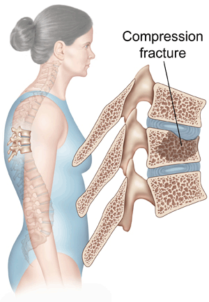
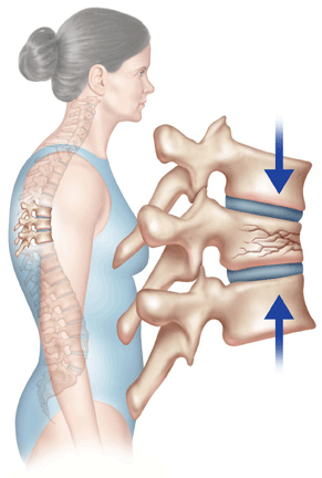
Perhaps the most common symptoms of osteoporosis are fractures-particularly vertebral compression fractures and hip fractures. The compression fractures in the spine that are caused by weakened vertebrae can lead to pain in the mid back. These fractures often stabilize by themselves and the pain eventually goes away. But the pain may persist if the crushed bone continues to move around and break.
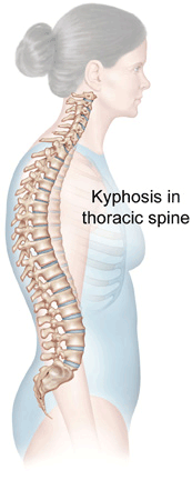
In severe cases of osteoporosis, actions as simple as bending forward can be enough to cause a "crush fracture" in a vertebra. This type of vertebral fracture causes loss of body height and a humped back, especially in elderly women. This disorder (called kyphosis) is an exaggeration in the curve of the mid back. It causes the shoulders to slump forward and the top of the back to look enlarged and humped.
Consult your doctor if you have symptoms of osteoporosis. Older women should discuss their risks with their physician, even if they are not currently showing any signs of osteoporosis. All women should be aware of the many preventive steps that can lower their risk of developing osteoporosis.
Learn about preventative measures for osteoporosis.
Diagnosis
Diagnosis generally begins with a physical exam that measures height, weight, and arm span. This gives a rough estimate of what your original height might have been in young adult life. Your posture and vertebral tenderness will also be checked.
Your doctor may ask you to have a bone mineral density test. Bone densitometry measures the density of your bone mass. This test is not part of a routine screening, but it will be done if osteoporosis is suspected or if you are at high risk for getting it. The test uses an X-ray beam to analyze bone density. The results are placed on a graph. A T-score shows how your bone density compares to the density of a healthy person who is 30 years old. Normal bone is between 0 and 1. Bones with T-scores between 1 and 2.5 are called osteopenic, meaning "too little bone." A T-score that is more than 2.5 below ideal levels indicates osteoporosis. Doctors also compare your scores to people your same age and sex. This is called a Z-score.
People at risk for osteoporosis can benefit by getting a bone mineral density test done earlier. The results can help identify if a problem exists so proper treatment can begin sooner.
Lab tests are conducted to rule out any secondary disorders that might be causing the osteoporosis. Tests of urine and serum are used to look for concentrations of calcium, serum protein, inorganic phosphorus, alkaline phosphates, or complete blood cell count (CBC). A CBC with a separate white cell count can be used to rule out other diseases. Biochemical measures of bone turnover and other clinical information can be considered. Elderly people should have thyroid function tests, serum, and urinary protein electrophoresis to rule out hyperthyroidism and multiple myeloma.
X-rays might be taken if your doctor suspects a fracture. An X-ray can also show if there are problems with bone content. An X-ray may detect problems with osteoporosis if the bones have lost 40 percent or more of their normal substance.
Treatment Options
There is still no cure for osteoporosis. But in recent years many effective treatments and prevention plans have been discovered. The best treatment for osteoporosis continues to be prevention.
Learn about preventative measures for osteoporosis.
Physical Therapy
Your doctor may have you work with a physical therapist. A well-rounded rehabilitation program assists in calming pain and inflammation, improving your mobility and strength, and helping you do your daily activities with greater ease and ability.
Physical therapists design treatment programs to improve your flexibility, strength, and posture. Exercises are chosen to help stabilize your spine while preventing bent positions of your spine. Therapists evaluate your balance and strength to make sure you are not at risk of having a fall. Therapy sessions may be scheduled two to three times each week for up to six weeks.
The goals of physical therapy are to help you
- Learn correct posture and body movements to counteract the effects of osteoporosis
- Perform a routine of safe weight bearing and resistance exercise
- Use safe lifting practices to avoid straining the spine
- Improve balance to prevent falling down
- Learn ways to manage your condition